Welcome to the Ford Lab
  • Home
  • RESEARCH
  • News
    • 2024-2025
    • 2023
    • 2022
    • 2021
    • 2020
    • 2019
    • 2018
    • 2017
  • People
    • Past Alumni
  • Publications
  • Positions Available
  • Contact
  • Links

Ford Lab Research

Image of lab benches inside Ford lab
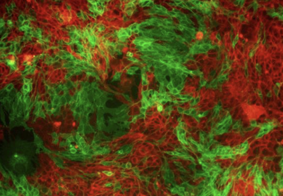
Picture of Tumor where Cells are undergoing EMT (green cells- undergoing EMT, red cells- epithelial)Picture of Tumor where Cells are undergoing EMT (green cells- undergoing EMT, red cells- epithelial)
Tumor Heterogeneity, Epithelial to Mesenchymal Transition (EMT), and Metastasis. We study the heterogeneity that arises in tumors when a subset of cells re-express developmental transcription factors (TFs) and undergo an EMT .  We are using biochemical and molecular approaches (mass spectrometry, RNAs-seq, and ChiP-seq), coupled with in vivo approaches to understand how crosstalk between tumor cells leads to enhanced metastasis, and how  TFs regulate metastasis cell autonomously and non-cell autonomously.  Figure to left shows breast cancer cells with a lineage tracing system where red is the epithelial population and green represents cells undergoing an EMT. 
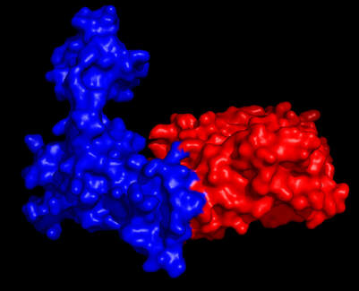
Structure of SIX1/EYA2 complexStructure of SIX1/EYA2 complex
Targeting Developmental TFs  to Inhibit Tumor Progression.  In collaboration with the Zhao laboratory in the Department of Biochemistry and Molecular Genetics, we have identified novel small molecules that can target the Six/Eya transcriptional complex .  The small molecules are currently being tested in cell based/animal models, and are anticipated to inhibit tumor growth and metastases while conferring limited side effects due to the paucity of expression of these developmental TFs in differentiated cells.  This project utilizes biophysical, biochemical, and structural approaches, as well as cell-based/animal model approaches.  Figure to left shows the SIX1/EYA domain crystal structure.

Tumor from mouse xenograft stained to identify tumor cells and various immune cellsTumor from mouse xenograft stained to identify tumor cells and various immune cells
Understanding how cancer cells evade the immune microenvironment.   Our laboratory is studying how the Eya Thr phosphatase acts, and how, when expressed in cancer cells, it contributes to the ability of the cells to avoid attack by the immune system.  Recent studies have focused on an Eya-Myc-PD-L1 axis that suppresses CD8+ cytotoxic T-cells, and ongoing studies examine other immune populations as well as how EMT contributes to tumor immune evasion.   Figure to left shows a Vectra Polaris stained tumor in which one can detect different immune populations.  ​
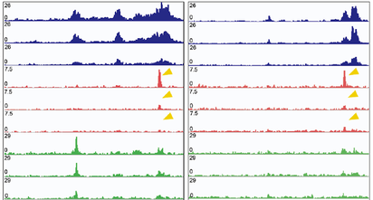

Picture of Chip-Seq tracksPicture of Chip-Seq tracks
Understanding the role of the Six/Eya transcriptional complex in pediatric tumors.  Using zebrafish (in collaboration with The Artinger Lab at UMN) as well as mouse models, we are studying how developmental transcription factors (TF) such as Six and Eya are regulated during normal embryogenesis, as well as how their dysregulation contributes to tumor progression in various pediatric malignancies ​including rhabdomyosarcoma, medulloblastoma, and Ewing sarcoma.  The figure to the left shows some Chip-Seq tracks.

Log2 fold changes in Ribosome protected fragments (Y-axis) vs RNA levels (X-axis) in normoxia vs hypoxiaLog2 fold changes in Ribosome protected fragments (Y-axis) vs RNA levels (X-axis) in normoxia vs hypoxia
Translational Control of Metastasis.  In this project, we are looking at how specialized translation in response to stresses (such as hypoxia, nutrient deprivation etc) influence metastasis, with a focus on the eIF3 translational complex.  This project is a collaboration with the Mukherjee and Zhao labs from Biochemistry and Molecular Genetics, and involves many different techniques such as ribo-seq, informatics analysis, 2D and 3D culture systems, drug development, and in vivo metastasis experiments.  The picture to the left is a plot showing transcriptional changes (X axis) when compared to translational changes (Y axis) one hour after a cellular stress. 

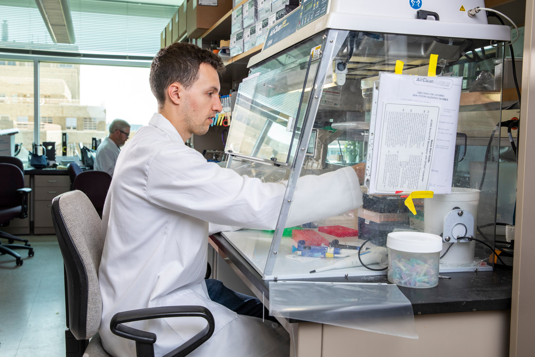
Connor busy at work at qPCR.
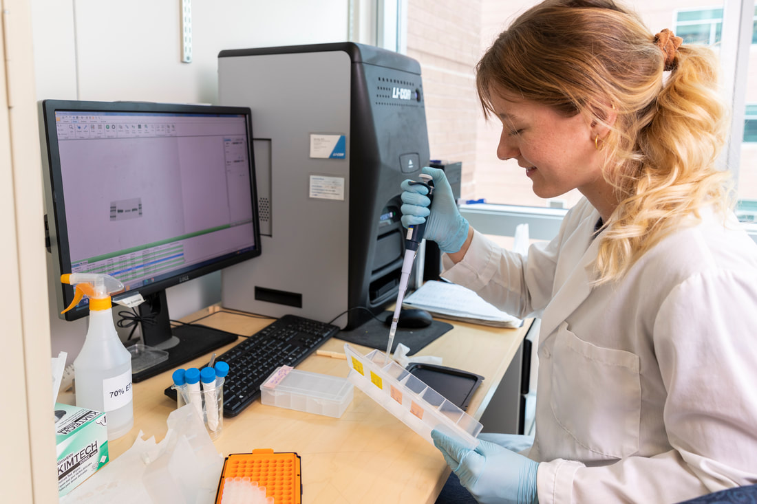
Kaiah developing her western blot experiments.
Picture
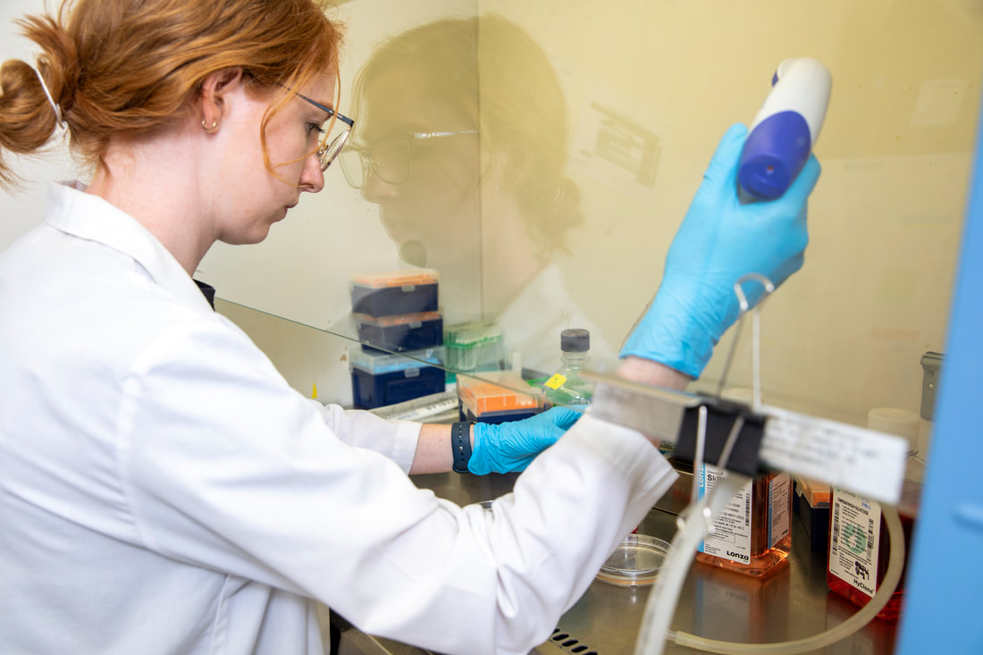
Annika at work in the hood on her project related to transcriptional and epigenetic regulation of RMS.
Picture
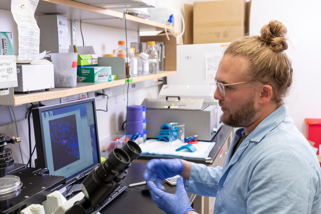
Art looking at some of his images in which he is examining alterations to mitotic spindles in GBM.
Proudly powered by Weebly
  • Home
  • RESEARCH
  • News
    • 2024-2025
    • 2023
    • 2022
    • 2021
    • 2020
    • 2019
    • 2018
    • 2017
  • People
    • Past Alumni
  • Publications
  • Positions Available
  • Contact
  • Links